Nervous system

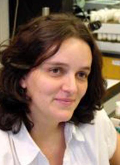
Amelia Gallitano

Professor, Basic Medical Sciences
Professor, Psychiatry
Professor, Neuroscience - GIDP
Member of the Graduate Faculty
Professor, BIO5 Institute
Contact
(602) 827-2131

Work Summary

The Gallitano Lab investigates the molecular mechanisms underlying the dual genetic and environmental risk for neuropsychiatric illnesses such as schizophrenia and mood disorders. We focus on immediate early genes that are activated in the brain by environmental stimuli, including stress, and regulate processes disrupted in mental illnesses. Ongoing studies examine how Egr3 regulates effectors including Arc and the serotonin 2A receptor to influence synaptic plasticity, memory, and behavior.

Research Interest

Amelia Gallitano, M.D., Ph.D. is an Associate Professor of Basic Medical Sciences and Psychiatry at the University of Arizona College of Medicine (UACOM) – Phoenix. Dr. Gallitano received her medical degree, and Ph.D. in Neuroscience, from The University of Pennsylvania School of Medicine. She completed residency training in Psychiatry at Columbia University, New York, and the New York State Psychiatric Institute, followed by a post-doctoral research fellowship at Washington University School of Medicine in St. Louis, where she was a faculty member in the Department of Psychiatry. She is a board-certified psychiatrist. Dr. Gallitano joined the UACOM - Phoenix as one of the founding faculty members in 2007, the year it opened to its first class of medical students. Research in her laboratory investigates how genes activated in the brain in response to stress may mediate the interaction between environment and genetic variations to influence the development of psychiatric illnesses such as schizophrenia and mood disorders. In addition to their molecular studies, the Gallitano Lab has recently initiated a translational research program to develop a biologically-based diagnostic test for schizophrenia emanating from their basic science findings. Since starting her laboratory, Dr. Gallitano has received numerous grants and awards, including one of the first National Institute of Health (NIH) R01 grants awarded to an Assistant Professor in the department of Basic Medical Sciences (BMS). Her laboratory has maintained continuous NIH funding since 2012. She is currently an Associate Professor with Tenure in the Department of BMS and Associate Professor of Psychiatry at the UACOM – Phoenix, and a member of the Interdisciplinary Graduate Program in Neuroscience. She is also a member of the Arizona State University Graduate Interdisciplinary Program in Neuroscience and an Adjunct Investigator at the Translational Genomics Research Institute. Her research focuses on investigating how immediate early genes may mediate the interaction of environmental stress and genetic predisposition to influence the development of psychiatric illnesses such as schizophrenia. Dr. Gallitano has published under the names: Gallitano, Gallitano-Mendel, and Mendel. Dr. Gallitano is also a co-founder and director of the UACOM-Phx Women in Medicine and Science Committee. She is committed to supporting the advancement of women and under-represented groups in the sciences and to mentoring trainees from all backgrounds who have a passion for neuroscience.

Daniela C Zarnescu

Professor, Cellular and Molecular Medicine
Professor, Neuroscience
Professor, Applied BioSciences - GIDP
Professor, Neurology
Professor, Genetics - GIDP
Professor, Molecular and Cellular Biology
Professor, Neuroscience - GIDP
Member of the Graduate Faculty
Professor, BIO5 Institute
Primary Department
Contact
(520) 626-1478

Work Summary

We are working to uncover the molecular mechanisms of aging and neurodegenerative diseases using a combination of genetic, computational and pharmacological tools, and a diverse array of experimental models. We also seek to develop therapies for ALS and related neurodegenerative diseases.

Research Interest

Fragile-X syndrome, which includes mental and physical defects and is the most common form of inherited mental retardation. Keywords: Neurodegeneration, ALS, Aging

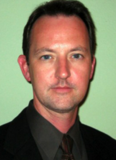
Russell S Witte

Professor, Medical Imaging
Professor, Biomedical Engineering
Professor, Applied Mathematics - GIDP
Professor, Neurosurgery
Professor, Optical Sciences
Professor, Neuroscience - GIDP
Professor, BIO5 Institute
Member of the General Faculty
Member of the Graduate Faculty
Primary Department
Department Affiliations
Contact
(520) 626-0346

Work Summary

We develop cutting-edge imaging technology, integrating light, ultrasound and electricity, to diagnose and treat diseases ranging from epilepsy to breast cancer. Novel sources for ultrasound contrast include optical and microwave absorption, mechanical strain, and electrical current. We visualize electrical brain “stormsˮ during uncontrollable seizures and envision “smartˮ photoacoustic agents that seek-and-destroy deadly tumors.

Research Interest

Dr. Russell Witte, a native Tucsonan, received a BS degree with honors in physics from the University of Arizona in Tucson (1993). Following travel abroad in Europe and Brazil, he began graduate studies in bioengineering at Arizona State University. His doctoral thesis (PhD, 2002) used chronic microelectrode arrays to describe sensory coding and learning-induced plasticity in the mammalian brain. He then moved to the University of Michigan in Ann Arbor and, as a post doc in the Biomedical Ultrasonics Laboratory, developed novel hybrid imaging techniques that integrate ultrasound, light, and/or microwaves for medical applications. In 2007, Dr. Witte returned to Tucson and is now Associate Professor of Medical Imaging, Optical Sciences and Biomedical Engineering at the University of Arizona. Dr. Witte’s Experimental Ultrasound and Neural Imaging Laboratory (EUNIL) devises cutting-edge imaging technology, integrating light, ultrasound and microwaves to diagnose and treat diseases ranging from chronic tendon disorders (tendinopathies) and irregular cardiac rhythms (arrhythmias) to breast cancer. By integrating different forms of energy, special effects are created that enable ultrasound imaging of optical absorption deep in tissue (photoacoustic imaging), mapping current source densities in the beating heart (acoustoelectric imaging), and elasticity imaging of human muscle and tendon for quantifying tissue mechanical properties. Dr. Witte's research further extends into nanotechnology and smart contrast agents, which have applications to functional brain imaging, cardiovascular disease, and cancer. Dr. Witte works closely with collaborators in the Colleges of Engineering, Optical Sciences and Medicine, as well as industry, to develop cutting-edge imaging technologies that potentially improve patient care. Dr. Witte is also a member of the Arizona Cancer Center, Sarver Heart Center and School of Mind, Brain, and Behavior, as well as the Neuroscience, Applied Mathematics, and Biomedical Engineering graduate interdisciplinary programs (GIDPs). Dr. Witte's vision is to develop a new generation of young investigators steeped in multiple disciplines branching from neuroscience, neural engineering, biochemistry, mathematics, biomedical imaging and, physics. He welcomes dreamers, brainstormers and problems solvers to join his team in search of the next great discovery in physics and medicine. Keywords: Biomedical Engineering/Medical Imaging

Jennifer A Teske

Associate Professor
Associate Professor, Neuroscience - GIDP
Associate Professor, Physiological Sciences - GIDP
Member of the General Faculty
Member of the Graduate Faculty
Primary Department
Department Affiliations
Contact
(520) 621-3081

Research Interest

Dr. Jennifer Teske, PhD is an Assistant Professor in the Department of Nutritional Sciences. Her primary research interest is the study of the metabolic consequences of environmental noise stress as it relates to the whole-organism stress response and human health.

Dianne K Patterson

Staff Scientist, Neuroimaging
Contact
(520) 621-1644

Work Summary

I analyze MRI images to understand more about how human language works. We use functional MRI to determine which brain regions are involved in different language tasks. We also look at diffusion MRI to learn about the quality of the wiring between regions.

Research Interest

I do neuroimaging, specifically fMRI and DTI. I am especially interested in brain networks and developments in neuroimaging software. We use independent component analysis to identify separate networks in the brain related to processing and learning language. My colleagues and I worked to improve fMRI analysis, display and data sharing options. Beginning with a web-based workbench designed for the dynamic exploration of map-based data, we worked to develop brain maps that could be similarly explored and demonstrated that this approach yielded results similar to those achieved by much more laborious and manual exploration techniques. This has improved our ability to streamline analyses, extract insights from our data and share data online. I have also worked on DWI analysis of the language system for the past 8 years. This has resulted in contributions to tract analyses (Wilson et al., 2011) and to the development of a novel technique (Patterson et al., 2015) to extract not only information about the properties of each tract but also information about the size and location of connected grey matter regions. We continue to explore the implications of these new measures. Keywords: fMRI, DWI, Language, Neuroimaging, MRI

Julie Elizabeth Miller

Associate Professor, Neuroscience
Associate Professor, Speech, Language, and Hearing Sciences
Primary Department

Work Summary

I am a neuroscientist who studies the impact of aging and neurodegenerative disease on voice and speech. My laboratory seeks a better understanding of the molecules, cells and circuits in the brain that support vocal production.

Research Interest

My laboratory studies neurogenetic mechanisms which underlie normal and abnormal motor speech using the zebra finch songbird. My particular focus is to investigate molecular and cellular pathways altered by speech disorders associated with natural aging and neurological diseases such as Parkinson’s Disease. To carry out these investigations, we use a combination of behavioral, genetic, biochemical and electrophysiological approaches that enable us to link changes at the molecular/cellular levels to alterations in neural circuits for birdsong/human speech. We also have collaborations with researchers working in mouse models to understand shared molecular pathway for vocal function. The end goal is to leverage the advantages offered by each species and an array of biological tools to further advance our understanding of how the brain controls vocalizations. Our laboratory website, including an updated publication list, can be found at: https://julieemiller.lab.arizona.edu/content/publications-abstracts

Aneta Kielar

Assistant Professor, Speech/Language and Hearing
Assistant Professor, Cognitive Science - GIDP
Assistant Professor, BIO5 Institute
Contact
(520) 621-1644

Work Summary

My research examines neural factors which affect language functions, and how these change across life-span and are influenced by stroke, brain injury and neurodegenerative disorders. In my work, I use combination of cognitive measures and multimodal neuroimaging techniques (fMRI, EEG/ERPs, MEG). I am also interested in recovery of function, and treatment approaches involving speech-language therapy in combination with noninvasive brain stimulation techniques.

Research Interest

My research program is centered on investigating the neurobiology of healthy language system, and changes in cognitive and language processing associated with stroke and neurological disorders. My interests include incorporating cognitive measures and multimodal neuroimaging methods, with a goal to understand the relationship between language and other aspects of cognition, as well as the neural dynamics related to brain damage, resilience, and recovery. My research efforts are directed towards identifying factors which affect language comprehension and production, and how these change with development and are influenced by aging, stroke, brain injury, and neurodegenerative disorders, including Primary Progressive Aphasia (PPA) and Alzheimer’s disease (AD). I study language processing at the multiple levels, using behavioral experiments and both structural (DTI, lesion-symptom mapping, voxel-based morphometry) and functional neuroimaging (fMRI, EEG, MEG). In addition, I am interested in neuroplasticity and application of noninvasive brain stimulation techniques (e.g., TMS, tDCS) to the treatment of aphasia and dementia. The long-term goal of my research is to understand the cognitive and neural processes that support recovery of cognitive and language functions after stroke. Keywords: stroke, aphasia, dementia, MRI, EEG, Language

Charles M Higgins

Associate Professor, Neuroscience
Associate Professor, Neuroscience - GIDP
Associate Professor, Applied Mathematics - GIDP
Associate Professor, Electrical and Computer Engineering
Associate Professor, Entomology / Insect Science - GIDP
Associate Professor, BIO5 Institute
Primary Department
Department Affiliations
Contact
(520) 621-6604

Research Interest

Charles Higgins, PhD, is an Associate Professor in the Department of Neuroscience with a dual appointment in Electrical Engineering at the University of Arizona where he is also leader of the Higgins Lab. Though he started his career as an electrical engineer, his fascination with the natural world has led him to study insect vision and visual processing, while also trying to meld together the worlds of robotics and biology. His research ranges from software simulations of brain circuits to interfacing live insect brains with robots, but his driving interest continues to be building truly intelligent machines.Dr. Higgins’ lab conducts research in areas that vary from computational neuroscience to biologically-inspired engineering. The unifying goal of all these projects is to understand the representations and computational architectures used by biological systems. These projects are conducted in close collaboration with neurobiology laboratories that perform anatomical, electrophysiological, and histological studies, mostly in insects.More than three years ago he captured news headlines when he and his lab team demonstrated a robot they built which was guided by the brain and eyes of a moth. The moth, immobilized inside a plastic tube, was mounted on a 6-inch-tall wheeled robot. When the moth moved its eyes to the right, the robot turned in that direction, proving brain-machine interaction. While the demonstration was effective, Charles soon went to work to overcome the difficulty the methodology presented in keeping the electrodes attached to the brain of the moth while the robot was in motion. This has led him to focus his work on another insect species.

Erika D Eggers

Associate Department Head, Research - Physiology
Member of the Graduate Faculty
Professor, BIO5 Institute
Professor, Biomedical Engineering
Professor, Neuroscience - GIDP
Professor, Physiological Sciences - GIDP
Professor, Physiology
Primary Department
Department Affiliations
Contact
(520) 626-7137

Work Summary

My laboratory studies how the retina takes visual information about the world and transmits it to the brain. We are trying to understand how this signaling responds to changing amounts of background light and becomes dysfunctional in diabetes.

Research Interest

The broad goal of research in our laboratory is to understand how inhibitory inputs influence neuronal signaling and sensory signal processing in the healthy and diabetic retina. Neurons in the brain receive inputs that are both excitatory, increasing neural activity, and inhibitory, decreasing neural activity. Inhibitory and excitatory inputs to neurons must be properly balanced and timed for correct neural signaling to occur. To study sensory inhibition we use the retina, a unique preparation which can be removed intact and can be activated physiologically, with light, in vitro. Thus using the retina as a model system, we can study how inhibitory synaptic physiology influences inhibition in visual processing. This intact system also allows us to determine the mechanisms of retinal damage in early diabetes. Keywords: neuroscience, diabetes, vision, electrophysiology, light

Thomas P Davis

Professor, Pharmacology
Professor, Pharmacology and Toxicology
Professor, Neuroscience - GIDP
Professor, Physiological Sciences - GIDP
Professor, BIO5 Institute
Primary Department
Department Affiliations
Contact
(951) 858-5720

Research Interest

Thomas Davis, PhD, and his lab continue its long-term CNS biodistribution research program, funded by NIH since 1981, by studying the mechanisms involved in delivering drugs across the blood-brain barrier to the C.N.S. during pathological disease states. Recently, Dr. Davis and his lab discovered specifica drug transporters which can be targeted to enhance delivery. They are also interested in studying the effect of hypoxia/aglycemia/inflammatory pain on endothelial cell permeability and structure at the blood-brain barrier. Dr. Davis has recently shown that short-term hypoxia/aglycemia leads to significant alterations in permeability which can be reversed by specific calcium channel antagonists. This work has significant consequences to the study of stroke. Additionally, he has discovered that peripheral pain has significant effects on BBB tight junction protein cytoarchitecture leading to variations in the delivery of analgesics to the CNS.